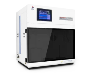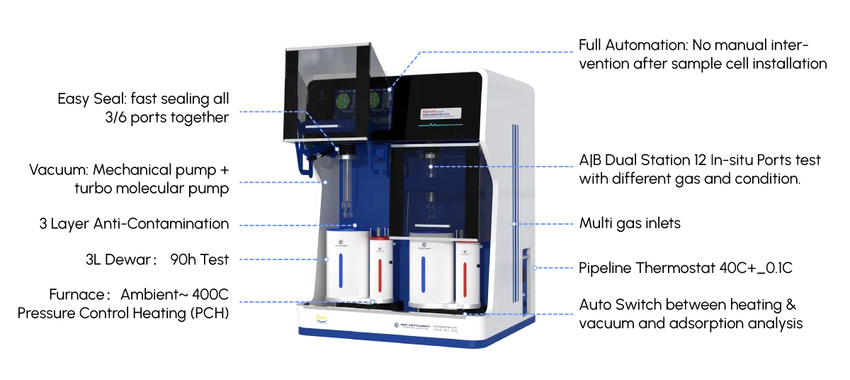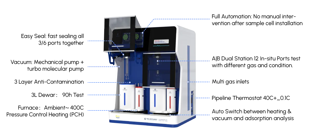
Integrating Machine Learning Algorithms with Micropore Analyzer Data for Rapid Pore Size Distribution Prediction

Abstract
Microporous materials play a pivotal role in energy storage, catalysis, gas separation, and environmental remediation. Accurate characterization of pore size distribution (PSD) is essential for understanding material performance; however, conventional methods—such as density functional theory (DFT) fitting and Monte Carlo simulations applied to physisorption isotherms—are computationally intensive and time-consuming. In this study, we propose a data-driven framework that integrates machine learning (ML) algorithms with micropore analyzer data to enable rapid and reliable PSD prediction. By leveraging experimental nitrogen adsorption–desorption isotherms collected from a diverse set of microporous samples, we trained and evaluated multiple ML models, including Random Forest, Support Vector Regression, Gradient Boosting Machines, and Artificial Neural Networks. Feature engineering was performed on raw isotherm data, pressure-step derivatives, and statistical descriptors to enhance model interpretability and predictive accuracy. The results demonstrate that the optimized ML models can predict PSD curves with high fidelity while reducing computation time by orders of magnitude compared to traditional DFT-based approaches. SHAP (SHapley Additive exPlanations) analysis further reveals the physical relevance of key features, bridging data-driven predictions with adsorption theory. This work highlights the potential of ML-assisted characterization workflows to accelerate materials screening and design, offering a practical pathway toward real-time PSD estimation in both research and industrial settings.

1. Introduction

Microporous materials—including zeolites, metal–organic frameworks (MOFs), activated carbons, and porous polymers—are widely used in applications ranging from molecular sieving and heterogeneous catalysis to carbon capture and energy storage [1–3]. A critical determinant of their performance is the pore size distribution (PSD), which governs diffusion kinetics, adsorption capacity, and selectivity. Consequently, accurate and efficient determination of PSD is indispensable in both fundamental research and industrial quality control.
Conventional PSD analysis typically relies on interpreting gas physisorption isotherms, most commonly nitrogen at 77 K. Classical approaches such as the Barrett–Joyner–Halenda (BJH) method, Horváth–Kawazoe (HK) model, and nonlocal/quasi-local density functional theory (NLDFT/QLDFT) have become standard in the field [4–6]. Among these, DFT-based methods are regarded as the most rigorous, accounting for fluid–fluid and solid–fluid interactions under realistic thermodynamic conditions. However, DFT calculations require significant computational resources and often involve iterative fitting procedures, limiting their utility in high-throughput characterization or real-time process monitoring.
In recent years, machine learning (ML) has emerged as a powerful tool in materials science, enabling rapid property prediction, inverse design, and automated data interpretation [7–9]. ML models excel at identifying complex, nonlinear relationships between measurable features and target properties, making them well suited for tasks where physics-based simulations are costly. Several studies have demonstrated successful ML applications in porosity-related problems, such as predicting Brunauer–Emmett–Teller (BET) surface areas from chemical composition or crystal structure [10,11]. Nevertheless, direct prediction of full PSD curves from experimental isotherm data remains underexplored, particularly in the micropore regime (<2 nm).
To address this gap, we present an integrated framework combining micropore analyzer data with supervised ML algorithms for rapid PSD prediction. Our approach bypasses the need for explicit DFT fitting by learning mappings from adsorption isotherms to PSDs using curated experimental datasets. We systematically compare multiple regression strategies, assess model generalizability across material classes, and employ explainable AI techniques to elucidate model behavior. The proposed methodology offers a scalable, low-latency alternative to conventional PSD analysis without sacrificing physical consistency.

2. Materials and Methods

2.1 Dataset Construction

Experimental nitrogen adsorption–desorption isotherms were compiled from published literature and in-house measurements, covering a broad spectrum of microporous materials: zeolites (e.g., ZSM-5, Y-type), MOFs (e.g., UiO-66, MIL-101), and activated carbons with varying degrees of activation. All samples were measured using commercial micropore analyzers (e.g., Micromeritics ASAP 2460) following standard degassing protocols. Relative pressure () ranged from to 0.99, with fine resolution in the low-pressure region () to resolve microporosity.
Ground-truth PSDs were obtained by applying NLDFT kernel models (e.g., N₂ at 77 K on carbon/silica) to the same isotherms using established software packages (e.g., MicroActive, Quantachrome TouchWin). This yielded continuous PSD curves discretized into 50–100 pore-width bins spanning 0.4–2.0 nm. A total of 850 validated samples were included, split into training (70%), validation (15%), and test (15%) sets.

2.2 Feature Engineering

Raw isotherm data were transformed into informative features to improve model learning:
  • Isotherm values​ at selected points (logarithmically spaced).
  • First and second derivatives​ of adsorption uptake with respect to , capturing inflection points associated with pore filling.
  • Statistical descriptors: area under the isotherm, hysteresis index, initial slope, and maximum uptake.
  • Textural parameters: BET surface area, total pore volume (), and micropore volume () calculated via standard methods.
All features were normalized to zero mean and unit variance prior to model training.

2.3 Machine Learning Models

Four representative ML algorithms were implemented and benchmarked:
  • Random Forest Regression (RFR):​ An ensemble of decision trees robust to noise and feature scaling.
  • Support Vector Regression (SVR):​ Using radial basis function kernels to capture nonlinearities.
  • Gradient Boosting Machines (GBM):​ Including XGBoost and LightGBM for efficient gradient-based optimization.
  • Artificial Neural Networks (ANN):​ Multilayer perceptrons with ReLU activations and dropout regularization.
Hyperparameters were tuned via grid search with 5-fold cross-validation on the training set. Model performance was evaluated using metrics including mean absolute error (MAE), root mean square error (RMSE), and coefficient of determination () computed over the entire PSD curve.

2.4 Explainability Analysis

SHAP values were employed to quantify feature contributions to model predictions, providing insights into which regions of the isotherm most strongly influence specific pore-size ranges. This facilitates physical interpretation and model trustworthiness.

3. Results and Discussion

3.1 Model Performance Comparison

Among the tested algorithms, ensemble methods (RFR and GBM) consistently outperformed SVR and ANN in terms of stability and accuracy. The best-performing GBM model achieved an average RMSE of 0.018 cm³/g across all pore-width bins and an value of 0.96 on the test set. Notably, prediction errors were lowest in the 0.5–1.2 nm range—critical for molecular sieving applications—and slightly higher near the micropore/mesopore boundary (1.8–2.0 nm), where capillary condensation effects complicate isotherm interpretation.
Compared to conventional NLDFT analysis, which requires several minutes per sample on a desktop workstation, the trained ML model generates a full PSD in milliseconds—a speedup exceeding three orders of magnitude. This enables near-real-time feedback during synthesis optimization or quality inspection.

3.2 Generalization Across Material Classes

When trained separately on carbonaceous versus crystalline materials, model accuracy improved significantly, suggesting that distinct adsorption mechanisms govern different material families. However, a unified model trained on mixed data still maintained acceptable performance (RMSE < 0.025 cm³/g), indicating robustness for general-purpose use. Transfer learning experiments further showed that pretraining on large synthetic datasets followed by fine-tuning on limited experimental data can mitigate data scarcity issues.

3.3 Physical Interpretability via SHAP Analysis

SHAP analysis revealed that low-pressure adsorption data () predominantly informed predictions for ultramicropores (<0.7 nm), consistent with monolayer adsorption and pore-filling theories. Mid-pressure features () correlated strongly with 0.7–1.5 nm pores, while derivative peaks aligned with known pore-filling transitions. These findings validate that the ML models internalize physically meaningful patterns rather than merely memorizing data.

3.4 Limitations and Future Directions

Despite promising results, several limitations remain. First, the current framework assumes N₂ at 77 K; extension to other adsorbates (e.g., Ar, CO₂) and temperatures will require additional training data. Second, highly disordered or hierarchical pores may introduce ambiguities not fully captured by integral isotherm features. Future work will explore convolutional neural networks to directly process isotherm curves as sequential data and incorporate structural priors (e.g., XRD patterns) to enhance predictive power.

4. Conclusion

This study demonstrates that integrating machine learning algorithms with micropore analyzer data enables rapid, accurate, and physically interpretable prediction of pore size distributions. By learning directly from experimental nitrogen adsorption isotherms, the proposed framework circumvents the computational bottlenecks of traditional DFT-based methods while preserving quantitative reliability. The approach is readily deployable in automated characterization pipelines and supports accelerated discovery and optimization of microporous materials. More broadly, this work exemplifies how data-driven techniques can complement—rather than replace—established physical models in materials characterization.

Gas Sorption vs. Mercury Intrusion: Which Pore Size Analyzer Technique Is Right for You?

Introduction

Characterizing the pore architecture of a material is fundamental to predicting its performance in applications ranging from catalysis and energy storage to pharmaceuticals and construction. Two of the most powerful and widely used techniques for this task are gas sorption (physisorption) and mercury intrusion porosimetry (MIP). While both aim to answer the same core question—”how big are the pores and how many are there?”—they operate on entirely different physical principles and probe vastly different regions of the pore size spectrum. Choosing the wrong method can lead to misleading data, wasted time, and flawed material designs. This article cuts through the technical jargon to provide a clear, decision-oriented comparison, helping you select the right tool based on your material type, pore size range, and the specific structural information you require.

The Fundamental Principle: Physisorption vs. Intrusion

At its core, the difference between these techniques is one of physical mechanism. Gas sorption relies on the physical adsorption of an inert gas (typically nitrogen, argon, or krypton) onto the surface of a material at cryogenic temperatures. As the relative pressure of the gas increases from near-zero to saturation, a multi-layer film forms on the pore walls, which eventually condenses into a liquid-like state within the pores via capillary condensation. The relationship between the pressure at which condensation occurs and the pore size is described by the Kelvin equation, allowing the calculation of pore size distributions down to the microporous regime.

Mercury intrusion, in contrast, is a high-pressure technique that forces a non-wetting liquid (mercury) into the pore structure. Because mercury does not spontaneously wet most materials due to its high surface tension, an external pressure is required to overcome the capillary resistance of the pores. The Washburn equation dictates that the pressure required to intrude mercury is inversely proportional to the pore radius; smaller pores require exponentially higher pressures. This fundamental distinction means that gas sorption is a gentle, non-destructive measurement, while mercury intrusion is an inherently destructive process that applies immense hydrostatic stress to the sample.

The Pore Size Domains They Cover

The single most important factor in your decision is the pore size range of interest. Gas sorption is the undisputed champion of the microporous (pores < 2 nm) and mesoporous (2–50 nm) regions. Using advanced models like Density Functional Theory (DFT) or Monte Carlo simulations, it can accurately resolve features down to the sub-nanometer scale, providing detailed information on micropore filling and surface heterogeneity. For materials like zeolites, metal-organic frameworks, or activated carbons, gas sorption is the only viable option among these two.

Mercury intrusion, however, shines in the macroporous range (pores > 50 nm) and can reliably characterize pores up to several hundred micrometers in diameter. While it can theoretically measure mesopores, the required pressures become impractically high (exceeding 400 MPa for pores under 4 nm) and risk crushing the sample or distorting the pore structure. Therefore, for materials with primarily micro- or small mesopores, gas sorption is mandatory. For macroporous solids such as concrete, soil, rocks, or large-pore foams, mercury porosimetry is the more suitable and practical choice.

Surface Area vs. Bulk Pore Volume

Consider what you truly need to know. Gas sorption is uniquely capable of providing a highly accurate specific surface area, most famously via the BET (Brunauer–Emmett–Teller) method. This is because the technique directly measures the amount of gas required to form a complete monolayer on the pore walls, offering a fundamental, surface-specific metric that is critical for understanding adsorption, catalysis, and reaction kinetics. The technique also delivers a comprehensive pore size distribution and total pore volume for mesopores.

Mercury intrusion, conversely, does not measure surface area directly. While it can produce an apparent surface area through the assumption of cylindrical pore geometry, this calculation is highly model-dependent and often unreliable for complex pore shapes. Its primary output is a bulk intrusion curve that yields total intrudable pore volume, bulk density, and an unambiguous pore throat size distribution. Crucially, MIP measures the entrance diameter of a pore, not its internal body. If a large internal cavity is accessed through a small neck, MIP will register the neck diameter, which can significantly underestimate the true internal pore size. Gas sorption, depending on the model, is more sensitive to the internal dimensions of the pore cavity itself.

Sample Integrity and Material Compatibility

The physical demands of each technique impose strict limitations on sample suitability. Gas sorption is a low-energy, low-temperature measurement that does not alter the sample in any way, provided that outgassing is performed correctly. It is safe for delicate frameworks, powdered catalysts, and organic materials. The only prerequisite is that the sample must be thoroughly degassed to remove volatile contaminants from the surface, a process that relies on heat and vacuum but does not mechanically stress the structure.

Mercury intrusion is a high-energy, high-pressure ordeal. The hydrostatic pressures required can exceed 200 MPa, which is sufficient to compact powders, fracture fragile frameworks, or plastically deform soft materials. This means that MIP results for compressible or brittle materials must be interpreted with extreme caution, as the intrusion curve may reflect mechanical deformation rather than true pore filling. Furthermore, the toxicity of mercury and the need for specialized handling equipment make this a more hazardous and costly procedure. For materials that are air- or moisture-sensitive, the mercury environment itself may cause unwanted chemical interactions.

Speed, Sample Size, and Practical Throughput

From a practical laboratory perspective, the operational differences are stark. Gas sorption is a relatively slow technique. A full nitrogen adsorption-desorption isotherm at cryogenic temperatures can take several hours to complete, especially if you include the necessary degassing step and multiple equilibration points for micropore analysis. This makes it less suitable for high-throughput quality control where rapid results are required.

Mercury intrusion, by contrast, is considerably faster. A typical analysis from vacuum to the maximum pressure and back down to atmospheric pressure can be completed in under an hour. This speed, combined with the fact that MIP requires minimal sample preparation (often just a dried piece of solid), makes it highly attractive for routine industrial testing and quality assurance. Additionally, MIP can analyze much larger sample pieces—often centimeter-scale chunks—providing a more representative bulk measurement of heterogeneous materials, whereas gas sorption typically requires a carefully weighed, homogeneous powder of a few hundred milligrams.

Interpreting the Data: Hysteresis and Connectivity

The information gleaned from each technique extends beyond just a number. The adsorption-desorption isotherm in gas sorption exhibits hysteresis for mesoporous materials, and the shape of this hysteresis loop provides invaluable qualitative information about pore geometry, such as whether the pores are cylindrical, ink-bottle shaped, or slit-like. This gives the researcher profound insight into the pore network’s topology and connectivity, aspects that are essential for diffusion and mass transport studies.

Mercury intrusion also exhibits hysteresis, but it arises from an entirely different phenomenon: the “ink-bottle” effect and contact angle hysteresis. The intrusion curve (high pressure) measures the pore throat size, while the extrusion curve (low pressure) provides information on pore body size. The extent of the hysteresis loop between intrusion and extrusion can directly indicate the degree of pore connectivity and the presence of constricted pore entrances. For applications like reservoir rock characterization, this connectivity information is often more valuable than the absolute pore size itself.

Making the Decision: A Practical Workflow

No single technique is universally superior. The optimal approach often involves using both methods sequentially to obtain a complete picture of a complex pore network. However, if a choice must be made, follow this logical workflow.

Start by asking what pore size region dominates your material. If your application involves molecular sieving, gas storage, or catalysis, and the critical dimensions are below 50 nanometers, you must use gas sorption. There is no substitute for its accuracy in the micro- and lower-mesopore range.

If your material is a dense ceramic, a rock core, a building material, or a foam with pore sizes exceeding 100 nanometers, mercury intrusion will provide faster, more robust, and more statistically representative data on pore throat distribution and bulk density.

If your material is fragile, expensive, or an organic polymer, strongly favor gas sorption to avoid irreversible damage during mercury pressurization.

Finally, consider your goal. If you need the absolute surface area to calculate catalytic activity, gas sorption is non-negotiable. If you need to know the critical pore throat size controlling fluid permeability, mercury intrusion offers a direct physical simulation of that process.

Conclusion

In summary, gas sorption and mercury intrusion are not competing alternatives but complementary tools suited for different ends of the pore size spectrum. Gas sorption is the technique of choice for precision surface area analysis and characterization of micropores and mesopores, providing detailed, non-destructive insights into pore geometry and surface energy. Mercury intrusion is the workhorse for macroporous materials, offering rapid, high-throughput analysis of bulk pore volume, density, and throat connectivity under realistic pressure conditions. By aligning your choice with your material’s pore size, mechanical robustness, and the specific physical property you need to measure, you can confidently select the technique that will deliver meaningful, actionable data for your research or quality control objectives.

Constant Pressure Adsorption of Water Vapor on Desiccants: Isotherms, Kinetics, and Regeneration Energy

  Solid desiccant dehumidification is a key technology for air conditioning, food preservation, and industrial drying. While traditional studies often focus on constant volume or gravimetric analysis, constant pressure adsorption offers a more realistic simulation of dynamic dehumidification processes (e.g., in a fixed-bed or rotary wheel). This paper examines the adsorption of water vapor on two common desiccants—Silica Gel (type RD) and Zeolite (13X)—under constant water vapor partial pressure. We analyze three critical aspects: equilibrium isotherms (capacity), kinetic behavior (rate of uptake), and the thermodynamic penalty of regeneration energy.
  1. Introduction
  Unlike vapor compression systems, desiccant systems rely on the affinity between water molecules and the desiccant surface. Under constant pressure conditions, the driving force for adsorption is the difference between the gas-phase partial pressure and the equilibrium pressure at the desiccant temperature. As adsorption progresses, the release of latent heat raises the desiccant temperature (thermal wave), complicating the isothermal assumption. This study maintains a constant external water vapor pressure (e.g., 1.5 kPa, equivalent to 50% RH at 25°C) to measure performance.
  2. Materials and Methods
  Desiccants: Silica Gel (mesoporous, mean pore dia. 2.5 nm) and Zeolite 13X (microporous, 0.7 nm).
  Setup: A constant pressure volumetric system (Sievert-type) with a water vapor reservoir maintained at fixed pressure using a feedback pressure regulator. Temperature was kept at 298 K for adsorption, and regeneration was studied from 323–473 K.
  Procedure: The sample was activated at 150°C under vacuum. Then, water vapor was introduced at constant pressure. Uptake was recorded by a microbalance, and the heat release was monitored via thermocouples.
  3. Adsorption Isotherms (Constant Pressure, 298 K)
  The uptake (g H₂O/g desiccant) versus time curves reach distinct plateaus depending on the desiccant type.
  Silica Gel exhibits a Type V isotherm. At low pressure (P < 0.5 kPa), minimal uptake is observed. However, at the target pressure of 1.5 kPa, it reaches a substantial capacity of approximately 0.35 g/g. This shape indicates multilayer formation followed by capillary condensation in the mesopores, providing a “gentle slope” that offers stable capacity across a range of relative humidity.
  In contrast, Zeolite 13X shows a Type I isotherm (Langmuir-like). At extremely low pressures (0.1 kPa), zeolite already absorbs about 0.20 g/g due to strong electrostatic fields within its micropores. At 1.5 kPa, it reaches approximately 0.28 g/g, but the curve flattens quickly thereafter. The key finding under constant pressure is that zeolite reaches its capacity much faster, yet its total capacity at 1.5 kPa is lower than that of silica gel due to the earlier completion of pore filling.
  4. Kinetics of Water Vapor Adsorption
  At constant pressure, the rate-limiting step was analyzed using the Linear Driving Force (LDF) model, revealing distinct temporal phases.
  During the early stage (t < 100 s), zeolite dominates. Its small pore openings create a high surface potential, capturing water rapidly. The rate constant (k_LDF) for zeolite is 0.045 s⁻¹, significantly higher than silica gel’s 0.012 s⁻¹. However, in the mid stage (100–1000 s), silica gel overtakes zeolite due to favorable macropore diffusion, where water molecules condense in mesopores without substantial resistance.
  A critical thermal effect complicates this picture. Constant pressure conditions exacerbate heat buildup because the released latent heat cannot dissipate quickly. Silica gel’s lower heat of adsorption (approximately 50 kJ/mol compared to zeolite’s 70 kJ/mol) means it stays cooler, thereby maintaining a higher driving force for continued adsorption. Conversely, zeolite heats up rapidly, reducing its kinetic advantage after about 200 seconds. The practical implication is clear: for rapid cycling applications (e.g., a rotary wheel at 15–30 rpm), zeolite performs better, whereas for deep dehumidification with slower cycles, silica gel is kinetically suitable.
  5. Regeneration Energy
  Regeneration is the reverse process at constant pressure but elevated temperature. The total energy (Q_total) required to remove 1 kg of water consists of sensible heat (heating the desiccant) and the isosteric heat of desorption.
  Silica gel requires a regeneration temperature range of 90–120°C. Its isosteric heat lies between 50 and 60 kJ/mol. Because water is held primarily by hydrogen bonding rather than strong ionic forces within its mesopores, the energy penalty is relatively low, around 2,800–3,200 kJ per kilogram of water removed. This makes silica gel suitable for use with waste heat or solar thermal energy.
  Zeolite 13X, however, demands much higher regeneration temperatures of 180–250°C for complete desorption. Its isosteric heat ranges from 70 to 85 kJ/mol, reflecting the deep potential wells within its micropores that hold water molecules tightly. Consequently, the energy penalty is substantially higher, reaching 6,500–8,000 kJ per kilogram of water. This necessitates high-grade heat from electric or gas combustion sources. The trade-off is that zeolite yields extremely dry air (dew point below -40°C). Furthermore, under constant pressure conditions, zeolite exhibits a pronounced hysteresis in regeneration—the desorption isotherm does not match the adsorption isotherm until the temperature is raised an additional 50°C. Silica gel shows minimal such hysteresis.
  6. Discussion: Trade-offs
  Comparing the two desiccants under constant pressure reveals a clear set of trade-offs. Silica gel offers a high capacity (0.35 g/g) at 1.5 kPa, a slow initial uptake rate, a low regeneration temperature (90–120°C), and low energy consumption per kilogram of water. This makes it best suited for air conditioning and crop drying applications. Zeolite 13X, on the other hand, provides a medium capacity (0.28 g/g), a fast initial uptake rate, but requires a high regeneration temperature (180–250°C) and high energy per kilogram of water. Its best use is in deep drying and cryogenic air preparation.
  A novel constant pressure analysis reveals that zeolite essentially wastes energy unless the application specifically requires very low dew points. For general HVAC purposes, silica gel’s lower regeneration energy outweighs its slower kinetics. Moreover, the constant pressure condition highlights that the thermal wave during adsorption significantly penalizes zeolite more than silica gel.
  7. Conclusion
  Constant pressure adsorption tests more accurately reflect real desiccant operation than static equilibrium tests. This study demonstrates three main points. First, silica gel achieves higher equilibrium capacity at 1.5 kPa due to capillary condensation in its mesopores. Second, zeolite exhibits faster kinetics initially but suffers from thermal self-inhibition under constant pressure, losing its advantage after a few hundred seconds. Third, the regeneration energy for zeolite is approximately 2.5 times higher than that for silica gel, making zeolite impractical for low-grade waste heat systems. Therefore, designers should match desiccant pore structure to the target partial pressure and cycle time. For energy-efficient dehumidification at constant pressure, hybrid desiccants (e.g., silica gel impregnated with hygroscopic salts) offer a promising middle ground.

Adsorption Isotherm Hysteresis: Causes and Implications for Porous Materials Characterization

  1. Introduction
  Adsorption isotherm are among the most fundamental tools in materials science for probing the surface area, pore size distribution, and porosity of solid materials. In many gas–solid adsorption experiments—especially those involving mesoporous solids—the desorption branch does not retrace the adsorption branch. This phenomenon, known as adsorption hysteresis, manifests as a loop on the isotherm plot and carries rich information about pore geometry, surface chemistry, and phase transitions within confined spaces. Understanding the origin and interpretation of hysteresis is therefore critical for accurate porous materials characterization.
  2. Definition and Classification of Hysteresis Loops
  Hysteresis loops are typically observed in the relative pressure range of approximately 0.4 < P/P₀ < 1.0, corresponding to capillary condensation in mesopores (2–50 nm). The IUPAC classifies hysteresis loops into several types:
  Type H1: Steep, nearly vertical adsorption and desorption branches; commonly associated with uniform cylindrical pores.
  Type H2: Wide loops with gradual desorption; often linked to complex, ink-bottle pore networks.
  Type H3 and H4: Characteristic of slit-shaped pores or layered structures, frequently found in plate-like particles or aggregated nanomaterials.
  These classifications provide a qualitative framework, but quantitative interpretation requires deeper insight into the underlying mechanisms.
  3. Physical Origins of Adsorption Hysteresis
  3.1 Capillary Condensation and Evaporation
  In mesopores, adsorbate molecules condense at pressures lower than the bulk saturation pressure due to enhanced intermolecular forces near the pore walls. The difference between adsorption (filling) and desorption (emptying) arises because evaporation is often hindered by metastable menisci or geometric constraints, leading to delayed emptying and thus hysteresis.
  3.2 Pore Geometry Effects
  The shape and connectivity of pores strongly influence hysteresis behavior:
  Cylindrical pores​ tend to produce narrow, reversible loops.
  Ink-bottle pores​ generate pronounced hysteresis because the narrow necks control evaporation while the wide bodies fill easily.
  Hierarchical pore networks​ introduce additional complexity through interconnected pathways and pore blocking.
  3.3 Surface Tension and Contact Angle Hysteresis
  Changes in surface tension and contact angle during adsorption and desorption cycles can shift equilibrium conditions. Surface heterogeneity, roughness, and chemical functional groups further modify wetting properties, contributing to loop asymmetry.
  3.4 Adsorbate Phase Transitions
  In some systems, structural transitions of the adsorbed phase (e.g., layering transitions, crystallization of confined fluids) occur only during one branch of the cycle, producing hysteresis even in the absence of geometric confinement.
  4. Implications for Porous Materials Characterization
  4.1 Pore Size Distribution Analysis
  Standard methods such as the Barrett–Joyner–Halenda (BJH) model rely on hysteresis data to calculate pore size distributions. However, misinterpretation of loop shapes can lead to erroneous pore dimensions if the assumed pore model does not match the real structure.
  4.2 Surface Area and Porosity Evaluation
  Hysteresis affects the determination of total pore volume and accessible surface area. For instance, incomplete desorption may underestimate surface area or overestimate microporosity when using models like BET (Brunauer–Emmett–Teller).
  4.3 Material Design and Application Relevance
  In catalysis, gas storage, and separation technologies, hysteresis provides indirect evidence of pore accessibility, transport resistance, and adsorption kinetics—factors that directly impact performance. Tailoring pore architecture to minimize undesirable hysteresis can enhance material efficiency.
  5. Limitations and Emerging Approaches
  Traditional interpretations of hysteresis loops often assume thermodynamic equilibrium and idealized geometries. In reality, kinetic effects, out-of-equilibrium states, and dynamic adsorption processes can dominate. Recent advances in in situimaging, molecular simulations, and time-resolved adsorption measurements are helping to decouple these contributions and refine pore structure analysis.
  6. Conclusion
  Adsorption isotherm hysteresis is far more than an experimental artifact; it encodes valuable information about pore morphology, surface interactions, and confined fluid behavior. A nuanced understanding of its causes enables more accurate characterization of porous materials and guides the rational design of advanced adsorbents, catalysts, and separation media. Future progress will depend on integrating classical thermodynamic models with modern experimental and computational techniques.

Step-by-Step Guide to Operating a BET Adsorption Analyzer for Accurate Results

BET Adsorption Analyzer method is the gold standard for determining the specific surface area of powders and porous materials. However, the accuracy of the results heavily depends on strict adherence to operational protocols, particularly regarding sample preparation and degassing. This guide outlines the standard operating procedure to ensure reliable and reproducible BET measurements.

1. Pre-Operation: Instrument Preparation & Calibration

Before handling samples, ensure the analyzer is ready for precise measurement:
  • Gas Supply:​ Turn on the nitrogen (adsorbate) and helium (for dead volume calibration, if applicable) cylinders. Adjust the regulator pressure to the manufacturer’s recommended working pressure (typically 0.1–0.2 MPa) .
  • Power Up:​ Switch on the vacuum pump, the analyzer main unit, and the control computer. Allow the thermal conductivity detector (TCD) or pressure transducers to warm up and stabilize (usually 30 minutes) .
  • System Check:​ Perform a leak check and calibrate the saturation pressure (). Verify the instrument’s accuracy using a certified reference material (CRM) with a known surface area (e.g., silica or alumina). The measured value should fall within of the certified value .

2. Sample Preparation

Accurate weighing and pretreatment are critical to avoid systematic errors:
  • Sample Mass:​ Weigh an appropriate amount of the dry sample into a clean sample tube. The mass depends on the estimated surface area:
    • High surface area (): 50–200 mg.
    • Low surface area (): 0.5–2 g.
    • Rule of thumb:Aim for a total surface area of 15–20 per tube to ensure a strong signal .
  • Particle Size:​ Ensure the sample is finely ground (particles ) to avoid internal mass transfer limitations, but avoid overly fine powders that may be lost during vacuuming .
  • Pre-weighing:​ Record the exact weight of the sample (tube + sample – tare weight) to a precision of at least 0.1 mg .

3. Sample Degassing (The Most Critical Step)

Degassing removes physisorbed water, solvents, and other contaminants from the pore surfaces. Incomplete degassing is the primary cause of inaccurate (usually low) BET results.
  • Mounting:​ Secure the sample tube (often with a glass wool plug or frit to prevent sample loss) into the degas station or a separate vacuum oven .
  • Parameter Setting:​ Set the degassing temperature and time.
    • Temperature:Must be below the sample’s phase transition or decomposition point (generally the melting point). Common ranges are 105–150°C for standard materials, up to 300°C for robust ceramics .
    • Time:Typically 2–6 hours (or overnight at lower temps) until the vacuum pressure stabilizes, indicating no more volatiles are evolving .
  • Execution:​ Apply vacuum and heat. Once completed, allow the sample to cool to room temperature under vacuumbefore venting slowly (usually with dry nitrogen or helium) to prevent immediate re-adsorption of ambient moisture .
  • Post-weighing (Optional but Recommended):​ Weigh the tube again after degassing to account for any mass loss (e.g., loss of volatiles), though many operators use the pre-degassing mass if the loss is negligible .

4. Analysis Setup

  • Transfer:​ Carefully transfer the degassed sample tube to the analysis port of the BET instrument. If using a filling rod (to reduce dead volume), insert it now. Ensure the seal is tight to prevent leaks .
  • Liquid Nitrogen:​ Fill the Dewar flask with liquid nitrogen (LN₂). Ensure the level is sufficient to submerge the sample bulb completely and remains stable throughout the run .
  • Software Configuration:​ Input the exact sample mass, select the adsorbate (N₂), and define the analysis parameters:
    • Relative Pressure Range ():​ For BET calculation, ensure points cover the 0.05–0.35 range (typically 5–7 points) .
    • Equilibrium Mode:​ Choose between continuous flow or fixed time intervals based on the sample’s adsorption kinetics (porous materials may require longer equilibrium times) .

5. Running the Test & Data Acquisition

  • Start the analysis via the software. The instrument will typically:
    1. Perform a dead volume measurement (often using He).
    2. Lower the LN₂ Dewar to immerse the sample (cooling to 77 K).
    3. Introduce doses of adsorbate gas at increasing values, measuring the equilibrated pressure to calculate adsorbed quantity.
    4. Optionally perform a desorption branch by decreasing .
  • Monitor the initial points to ensure the system is stable and no leaks are present.

6. Data Processing & Validation

Once the isotherm is generated:
  • BET Plot:​ The software automatically plots vs. .
  • Linearity Check:​ Select the linear region (usually ). A valid BET analysis requires a correlation coefficient () (or per some stricter SOPs). If is low, the data or the selected range may be invalid .
  • Calculation:​ The software calculates the monolayer adsorbed volume () from the slope and intercept, and subsequently the specific surface area () using the formula:
    Where is Avogadro’s number, is the cross-sectional area of N₂ (0.162 nm²), is the molar volume, and is the sample mass .

7. Shutdown Procedure

  • Remove the sample tube and Dewar. Allow the sample tube to warm to room temperature before opening to avoid condensation.
  • Clean the sample tube thoroughly (wash with solvent, dry, or bake out) for the next use .
  • Close the gas cylinder valves, shut down the software, turn off the instrument, and finally the vacuum pump.
  • Record the usage in the logbook .

The Role of Porous Structures in Gas Adsorption Processes

Abstract

Porous structures play a pivotal role in gas adsorption processes, which are crucial for various industrial applications such as gas storage, separation, and purification. This article comprehensively explores the significance of porous structures in gas adsorption, including different types of porous materials, their characteristics, adsorption mechanisms, and recent research trends.

1. Introduction

Gas adsorption is a fundamental process with wide – ranging applications in environmental protection, energy storage, and chemical engineering. Porous materials, with their unique structural features, offer high surface areas and well – defined pore networks that are essential for efficient gas adsorption. The ability to control and optimize the porous structure of materials is key to enhancing adsorption performance.

2. Types of Porous Structures

2.1 Porous Carbon Materials

Porous carbon materials are a diverse family with different dimensional structures. Zero – dimensional (0D) types include carbon quantum dots, fullerenes, and carbon nanospheres. One – dimensional (1D) forms are carbon fibers, carbon nanotubes, and carbon nanowires. Two – dimensional (2D) configurations consist of graphene and graphdiyne, and three – dimensional (3D) architectures include diamond, graphite, activated carbon, carbon molecular sieves, carbon foams, and carbon aerogels. These materials are characterized by unique porous structures, high surface areas, abundant microporosity, and chemical stability, making them suitable for gas adsorption and storage .

2.2 Metal – Organic Frameworks (MOFs)

MOFs are a class of porous materials with high designability. They are composed of metal nodes and organic ligands, which can be tuned to achieve different pore sizes, shapes, and surface functionalities. MOFs have shown great potential in gas storage, such as hydrogen and methane storage, as well as selective gas adsorption. For example, PCN – 14 has demonstrated high methane uptake capacity, exceeding the US DOE target for methane storage .

2.3 Porous Coordination Cages (PCCs)

PCCs, also known as metal – organic cages (MOCs) or metal – organic polyhedra (MOPs), have discrete cage – like architectures and permanent cavities. They are assembled through weak interactions such as H – bonds, van der Waals forces, and π – π stacking. PCCs can be designed to have both intrinsic pores from cage cavities and extrinsic voids from loose molecular packing, enabling selective gas adsorption and separation .

2.4 Three – Dimensional Ordered Macropores (3DOM)

3DOM catalysts have highly ordered macroscopic pore structures. These structures provide large specific surface areas and reduce mass transfer resistance, promoting gas molecule diffusion and adsorption. They are being studied for applications in gas purification, such as the removal of volatile organic compounds (VOCs), CO, NOx, CO2, and H2S .

3. Characteristics of Porous Structures Affecting Gas Adsorption

3.1 Surface Area

A high surface area is generally associated with more adsorption sites, which can enhance the adsorption capacity. For example, MOFs can have extremely high Brunauer – Emmett – Teller (BET) surface areas, up to 3800 m²/g in some cases, leading to high gas uptake capacities .

3.2 Pore Size and Distribution

The pore size needs to be appropriately matched with the size of the gas molecules for effective adsorption. Micropores (pore size < 2 nm) are often crucial for gas storage as they can provide strong gas – solid interactions. Mesopores (2 – 50 nm) can facilitate mass transfer, and a hierarchical porous structure with a combination of micro – and mesopores can optimize both adsorption capacity and kinetics. In direct air capture systems, a balanced pore network of mesopores and micropores has been shown to yield high adsorption efficiency .

3.3 Surface Chemistry

The surface chemistry of porous materials can be modified to enhance selectivity towards specific gases. For instance, in silica – based porous materials, surface functionalization can adjust the properties for better gas adsorption. Also, in porphyrin – based porous materials, varying the coordinated metal cation can modulate gas adsorption selectivity .

4. Adsorption Mechanisms in Porous Structures

4.1 Physisorption

Physisorption occurs through weak van der Waals forces between the gas molecules and the pore walls. It is a reversible process and is often dominant at low temperatures. In nanopores, gas adsorption can occur in different layers near the pore wall, such as the absorption layer adjacent to the pore wall, the Knudsen layer where diffusion is influenced by the absorption layer, and the bulk layer where gas – gas interactions dominate .

4.2 Chemisorption

Chemisorption involves a chemical reaction between the gas molecules and the adsorbent surface. It is usually stronger and more selective than physisorption. For example, in gas purification processes, chemisorption can be used to selectively remove specific pollutants by forming chemical bonds with the adsorbent .

5. Recent Research Trends

Recent research has focused on developing more efficient and selective porous materials for gas adsorption. For example, in porous tetrapyrrolic materials, modulating the central metal cation can improve gas uptake selectivity, such as Co – OX1 showing improved CO2 uptake . Also, in the field of MOFs, efforts are being made to improve their stability, especially in the presence of water, for practical applications .

6. Conclusion

Porous structures are indispensable in gas adsorption processes. Different types of porous materials, such as porous carbon, MOFs, PCCs, and 3DOM, offer unique advantages in terms of surface area, pore size, and surface chemistry. Understanding the adsorption mechanisms and continuously optimizing the porous structure through research will lead to more efficient gas adsorption technologies for various applications, including environmental protection and energy storage.

In Situ Characterization of Gas Adsorption Mechanisms in Hierarchical Zeolites

Abstract:
Hierarchical zeolites, which integrate microporosity with meso- or macroporosity, offer enhanced mass transport and improved accessibility to active sites compared to conventional microporous zeolites. Understanding gas adsorption mechanisms in these complex structures is crucial for optimizing their performance in gas separation, storage, and catalysis. This article reviews recent advances in the in situ characterization of gas adsorption processes within hierarchical zeolites. By employing techniques such as in situ infrared (IR) spectroscopy, in situ Raman spectroscopy, and in situ small-angle X-ray scattering (SAXS), researchers can now probe the dynamic behavior of adsorbed molecules at the molecular level. Key mechanisms discussed include: (i) sequential pore filling in micro- and mesopores, (ii) surface-mediated phase transitions, (iii) confinement effects on adsorbate structure, and (iv) cooperative adsorption between different pore regimes. These insights reveal how hierarchical architectures alter adsorption thermodynamics and kinetics, leading to improved selectivity and capacity. The article concludes with perspectives on emerging multimodal in situ approaches and their potential to guide the rational design of hierarchical zeolites for targeted gas adsorption applications.

1. Introduction

Zeolites are crystalline aluminosilicates with well-defined micropores (typically <2 nm), which grant them molecular sieving properties and high surface areas. However, their exclusive microporosity often leads to diffusion limitations, particularly for bulky molecules, reducing the utilization of internal active sites. Hierarchical zeolites address this limitation by introducing secondary porosity (mesopores of 2–50 nm and/or macropores >50 nm), creating a multi-level pore network that facilitates faster mass transport while retaining the intrinsic microporous framework.

Gas adsorption in hierarchical zeolites is not simply a superposition of micro- and mesopore behavior. Instead, the proximity and connectivity between pore regimes give rise to synergistic effects. For instance, mesopores can act as reservoirs or transport channels that feed molecules into micropores, while micropores can impose selectivity on the overall adsorption process. To unravel these complex mechanisms, in situ characterization—measuring adsorption under realistic conditions (pressure, temperature, gas composition)—is essential. Unlike ex situ methods, in situ techniques capture transient states, intermediate species, and structural rearrangements during gas uptake.

2. Key In Situ Characterization Techniques

2.1 In Situ Infrared (IR) Spectroscopy
In situ IR spectroscopy monitors vibrational modes of adsorbed molecules and zeolite framework functional groups (e.g., silanols, Brønsted acid sites) as a function of gas pressure or time. For hierarchical zeolites, IR can distinguish between molecules adsorbed in micropores (confined, often perturbed bands) versus mesopores (more bulk-like bands). The shift in C–O or O–H stretching frequencies, for example, reveals hydrogen bonding interactions with pore walls. Time-resolved IR further enables tracking of site competition: gas molecules first occupy high-energy micropores, followed by weaker mesopore adsorption sites.

2.2 In Situ Raman Spectroscopy
Complementary to IR, Raman spectroscopy is particularly sensitive to non-polar adsorbates and symmetric vibrations. In hierarchical zeolites, in situ Raman can probe the formation of molecular clusters or condensed phases within mesopores. For instance, during CO₂ or CH₄ adsorption, the appearance of lattice modes in the Raman spectrum signals pore confinement effects. Spatially resolved Raman (confocal Raman microscopy) has been used to map adsorbate distribution across different pore regimes in individual zeolite crystals.

2.3 In Situ Small-Angle X-ray Scattering (SAXS)
SAXS provides information on electron density variations, making it ideal for studying gas-induced changes in pore filling. In situ SAXS, combined with adsorption isotherms, can quantify the fraction of mesopores filled at a given relative pressure. Anomalous SAXS (ASAXS) using contrast variation (e.g., tuning X‑ray energy near absorption edges of probe molecules like Xe or Kr) allows selective visualization of gas density within pores. This technique has revealed that in hierarchical zeolites, mesopores often fill before micropores—a reverse order compared to purely microporous materials—due to capillary condensation in mesopores.

2.4 In Situ Neutron Scattering
Neutrons are highly sensitive to light elements (H, C, O) and can penetrate high-pressure cells. In situ neutron diffraction and quasielastic neutron scattering (QENS) elucidate the positions and mobilities of adsorbed molecules. For hierarchical zeolites, QENS has demonstrated that translational diffusion in mesopores is orders of magnitude faster than in micropores, confirming the “highway” role of mesopores.

3. Adsorption Mechanisms Revealed by In Situ Studies

3.1 Sequential and Cooperative Pore Filling
Classical models assume independent adsorption in micro- and mesopores. However, in situ IR and SAXS show a more nuanced picture: at low relative pressures, micropores fill first due to strong adsorbent–adsorbate interactions. Once micropores saturate, mesopores begin to fill via multilayer adsorption and capillary condensation. In hierarchical zeolites, the transition between these regimes is often smoother than in physical mixtures of micro- and mesoporous materials, indicating a cooperative effect. For example, the mesopore wall provides a “pre-layer” of adsorbed molecules that can migrate into adjacent micropores through pore mouths, maintaining a near-equilibrium chemical potential across the hierarchy.

3.2 Confinement-Induced Structural Changes
In micropores, molecules are forced into specific orientations or even distorted conformations. In situ Raman has identified that benzene adsorbed in the micropores of hierarchical ZSM‑5 exhibits a red shift of its ring-breathing mode compared to liquid benzene, indicating strong confinement. In mesopores (e.g., 10–20 nm diameter), confinement is weaker but still sufficient to induce layering near the pore wall. This layering has been directly imaged by in situ SAXS through oscillatory scattering patterns.

3.3 Surface-Mediated Phase Transitions
Capillary condensation in mesopores is a first-order phase transition that depends on pore diameter, surface chemistry, and temperature. In situ SAXS hysteresis loops reveal that hierarchical zeolites often exhibit a reduced hysteresis width compared to ordered mesoporous silicas, due to the presence of micropores that act as “nucleation sites” for condensation. Furthermore, in situ IR has shown that in the presence of polar surface groups (e.g., silanols), water or alcohol adsorption proceeds via cluster formation rather than continuous film growth, altering the condensation pressure.

3.4 Competitive and Selective Adsorption in Mixtures
For gas mixtures (e.g., CO₂/N₂, CH₄/N₂), in situ IR combined with mass spectrometry allows real-time monitoring of composition changes in the adsorbed phase. In hierarchical zeolites, the selectivity is not simply a function of micropore size. Mesopores can pre-concentrate the more polarizable component (e.g., CO₂) through weaker van der Waals forces, delivering a higher local concentration to micropore entrances. This “antenna effect” enhances overall selectivity and capacity, as demonstrated for CO₂ capture using hierarchical zeolite 13X.

4. Case Study: CO₂ Adsorption in Hierarchical ZSM‑5

To illustrate the power of in situ characterization, consider the adsorption of CO₂ in hierarchical ZSM‑5 prepared by desilication. In situ IR at 298 K reveals: (i) at P < 0.01 bar, asymmetric stretching of CO₂ appears at 2342 cm⁻¹, characteristic of adsorption on extra-framework Al species in micropores; (ii) as P increases to 0.1 bar, a second band at 2335 cm⁻¹ emerges, assigned to CO₂ in mesopores; (iii) simultaneously, the bending mode of zeolite framework (550 cm⁻¹) shifts, indicating lattice relaxation due to adsorbate stress. In situ SAXS shows that mesopores start filling at P/P₀ ≈ 0.2, but the micropores continue to uptake CO₂ until P/P₀ ≈ 0.6, revealing that the two regimes operate in parallel rather than sequentially. Neutron diffraction further locates CO₂ molecules preferentially near Al-rich regions in the micropores and at silanol nests in mesopore walls.

5. Challenges and Future Directions

Despite progress, several challenges remain. First, time resolution: many in situ techniques trade off temporal vs. spatial resolution. Fast processes (seconds) require synchrotron or neutron sources with high flux. Second, sample heterogeneity: hierarchical zeolites often contain a distribution of pore sizes; in situ methods need to disentangle contributions from different pores. Third, operando conditions: combining adsorption measurements with catalytic reaction (operando) is still rare for hierarchical zeolites.

Future developments include:

  • Multimodal in situ cells: Simultaneous IR + SAXS or Raman + neutron diffraction on the same sample under identical conditions.

  • Machine learning-assisted data analysis: Extracting component-specific signals from complex, overlapping spectra.

  • In situ electron microscopy: Environmental TEM (ETEM) with differential pumping to image gas adsorption at the single-particle level, though careful electron beam effects must be mitigated.

6. Conclusion

In situ characterization has transformed our understanding of gas adsorption in hierarchical zeolites. Far from being a simple combination of independent pore systems, these materials exhibit cooperative mechanisms—sequential filling, confinement-induced structuring, surface-mediated condensation, and antenna effects—that arise from the intimate coupling between micro- and mesopores. Techniques such as in situ IR, Raman, SAXS, and neutron scattering provide complementary windows into the molecular behavior of adsorbates under realistic conditions. The insights gained are already guiding the rational design of hierarchical zeolites with tailored pore architectures for energy-efficient gas separation, carbon capture, and hydrocarbon storage. As in situ methods become faster, more sensitive, and easier to combine, we can expect a new era of precision engineering of hierarchical nanoporous solids.

Recent Innovations in BET Surface Area Analyzer Technology

Recent Innovations in BET Surface Area Analyzer Technology

The Brunauer-Emmett-Teller (BET) theory remains the cornerstone for determining the specific surface area of porous materials through nitrogen adsorption measurements. While the fundamental principles endure, recent technological advancements have dramatically enhanced the precision, speed, accessibility, and applicability of BET surface area analyzer across diverse scientific and industrial fields.
1. Enhanced Automation and Workflow Integration:
Modern instruments increasingly feature sophisticated automation capabilities. Robotic sample loading systems minimize manual intervention, reducing operator error and enabling high-throughput analysis – crucial for quality control in industries like catalysts or pharmaceuticals. Integrated software now often includes automated degassing protocols, optimized equilibration criteria, and intelligent data reduction routines that guide users through complex multi-point BET calculations, significantly improving reproducibility and efficiency.
2. Improved Sensitivity and Accuracy:
Detector technology has seen significant upgrades. Advanced pressure transducers with higher resolution and stability allow for more precise measurement of low-pressure adsorption isotherms, critical for accurately characterizing ultra-microporous materials (pore width < 0.7 nm). Furthermore, refined dosing algorithms and improved vacuum systems ensure better baseline stability and faster attainment of equilibrium, leading to more reliable surface area values, especially for challenging samples with low surface areas or slow adsorption kinetics.
3. Expanded Pore Size Analysis Capabilities:
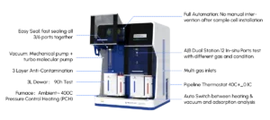
While BET focuses on monolayer formation, modern analyzers seamlessly integrate advanced physisorption models within the same instrument. Real-time calculation and visualization of pore size distributions using methods like Density Functional Theory (DFT) and Non-Local Density Functional Theory (NLDFT) are now standard features. This allows researchers to simultaneously obtain surface area anddetailed pore structure information (micro-, meso-, macropores) from a single experiment, providing a more complete material characterization.
4. Miniaturization and Portability:
A notable trend is the development of compact, benchtop, and even portable BET analyzers. These utilize novel sensor technologies (e.g., MEMS-based pressure sensors) and simplified gas handling systems. While potentially sacrificing some absolute accuracy compared to high-end lab systems, they offer rapid screening capabilities, field-deployable analysis (e.g., soil science, mineral exploration), and reduced cost of ownership, democratizing access to surface area measurements.
5. User-Centric Software and Data Management:
Software interfaces have evolved into powerful, intuitive platforms. Features include:
  • Automated Quality Checks:​ Flagging non-compliant data points based on IUPAC guidelines or user-defined criteria.
  • Advanced Visualization:​ Interactive plotting of adsorption/desorption isotherms and pore distribution curves.
  • Cloud Connectivity & LIMS Integration:​ Facilitating secure data storage, remote monitoring, collaboration, and seamless integration into laboratory workflows.
  • Predictive Modeling Tools:​ Some platforms now incorporate AI/ML algorithms to suggest optimal experimental parameters or predict properties based on historical data.
Impact and Future Directions:
These innovations collectively empower researchers and manufacturers to characterize increasingly complex materials – from metal-organic frameworks (MOFs) and graphene derivatives to battery electrodes and pharmaceutical excipients – with unprecedented ease and confidence. The drive towards faster analysis (<30 mins for routine samples), lower detection limits (approaching 0.01 m²/g), and seamless integration with other characterization techniques (e.g., TGA, XRD) continues. Looking ahead, further miniaturization, enhanced automation for complex sample types (powders, films, fibers), and deeper integration of computational modeling directly within the analyzer workflow promise to solidify BET analysis as an even more indispensable tool in materials discovery and process optimization.

Pulse Titration as a Tool for Rapid Metal Ion Quantification

Introduction

Pulse titration is an electroanalytical technique that combines the principles of potentiometry​ and chronoamperometry​ to enable rapid, sensitive, and selective quantification of metal ions in solution. Unlike traditional titrations (e.g., EDTA complexation or precipitation), pulse titration uses short, controlled potential pulses to drive redox reactions or complexation events, with current transients measured at each step. This method is particularly valuable for high-throughput analysis, on-site monitoring, and samples with low analyte concentrations, where speed and minimal sample preparation are critical.

Core Principles

The technique relies on two key components:
  1. Pulsed Potential Application: A working electrode (e.g., glassy carbon, gold) is subjected to a sequence of potential steps (pulses) over time. Each pulse is designed to either:
    • Oxidize/reduce the target metal ion (e.g., at a reducing potential).
    • Induce complexation with a ligand (e.g., EDTA) by adjusting the potential to favor binding.
  2. Current Transient Analysis: The current response to each pulse is recorded. The current magnitude depends on the concentration of the electroactive species (metal ions or their complexes) in the diffusion layer. By correlating current changes with known standards, the metal ion concentration is determined.
A critical advantage is the use of short pulse durations​ (milliseconds to seconds), which minimizes side reactions (e.g., double-layer charging) and enhances signal-to-noise ratios compared to steady-state methods.

Experimental Setup

A typical pulse titration system includes:
  • Three-Electrode Cell: Working electrode (sensing surface), reference electrode (e.g., Ag/AgCl), and counter electrode (e.g., platinum wire).
  • Potentiostat: Generates precise potential pulses and measures current responses.
  • Software: Controls pulse parameters (amplitude, duration, frequency) and analyzes data (e.g., peak current vs. concentration calibration curves).
Common modifications include using modified electrodes (e.g., with chelating agents like 8-hydroxyquinoline) to improve selectivity for specific metal ions.

Key Advantages Over Traditional Methods

  1. Speed: Each titration step takes seconds, enabling results in minutes—far faster than manual titrations (30+ minutes) or even some automated systems.
  2. Sensitivity: Detection limits can reach sub-micromolar levels (to nM) due to the enhanced current response from pulsed potentials.
  3. Selectivity: By tuning pulse potentials, interference from coexisting ions (e.g., in analysis) is reduced. For example, a potential pulse specific to reduction avoids oxidizing .
  4. Minimal Sample Preparation: Works with turbid or colored samples (unlike spectrophotometry) and requires no complex pre-treatment (e.g., digestion).
  5. Automation Compatibility: Easily integrated with flow systems for continuous monitoring (e.g., industrial wastewater streams).

Applications

Pulse titration is widely used in environmental, clinical, and industrial settings:
  • Environmental Monitoring: Rapid detection of heavy metals (, , ) in water and soil extracts. For example, it has been used to quantify in river water with a detection limit of 0.1 in <10 minutes.
  • Clinical Chemistry: Analysis of trace metals in blood (e.g., , ) for diagnosing deficiencies or toxicities.
  • Industrial Quality Control: Monitoring metal ion concentrations in plating baths (e.g., , ) or pharmaceutical formulations.

Limitations and Challenges

  • Electrode Fouling: Adsorption of metal ions or organic matter on the working electrode can degrade performance over time. Regular cleaning (e.g., with acid rinses) or disposable electrodes mitigate this.
  • Matrix Effects: High ionic strength or presence of surfactants may alter current responses. Calibration with matrix-matched standards is necessary.
  • Instrument Cost: Potentiostats with pulse capabilities are more expensive than basic titrators, though costs are decreasing with technological advances.

Recent Developments

Advancements in nanomaterials​ (e.g., graphene-modified electrodes) have improved sensitivity and anti-fouling properties. Additionally, machine learning​ integration now enables real-time data processing, further reducing analysis time. For example, a 2023 study used a machine learning algorithm to predict concentrations from pulse current data with 95% accuracy, cutting post-processing time by 50%.

Conclusion

Pulse titration represents a powerful evolution of traditional titration, offering unmatched speed, sensitivity, and automation for metal ion quantification. As miniaturization and smart sensor technologies advance, it is poised to become a standard tool in point-of-care diagnostics, environmental field testing, and industrial process control.

How Pore Size Analyzers Enable Breakthroughs in Catalyst and Battery Research

How Pore Size Analyzers Enable Breakthroughs in Catalyst and Battery Research
In the quest to engineer materials that power cleaner energy, faster chemical reactions, and longer-lasting technologies, understanding the microscopic architecture of porous substances has emerged as a linchpin of innovation. At the heart of this pursuit lies pore size analysis—a suite of techniques that maps the distribution, volume, and connectivity of pores within materials. Far from being mere measurement tools, pore size analyzers have become indispensable partners in advancing catalyst and battery research, unlocking breakthroughs that redefine what’s possible in these fields.

Decoding Porosity: The Unsung Hero of Function

Porous materials are not just empty spaces; they are dynamic landscapes where interactions between molecules, ions, and electrons unfold. In catalysts, pores act as nanoscopic reactors, controlling how reactant molecules access active sites and how products diffuse away. A catalyst with poorly tuned pore sizes might trap reactants or block product release, crippling efficiency. Similarly, in batteries, pores govern ion transport: too narrow, and ions face tortuous paths that slow charging; too wide, and the material may lack the surface area needed for stable energy storage. Pore size analyzers—ranging from gas adsorption-based methods like BET (Brunauer-Emmett-Teller) and BJH (Barrett-Joyner-Halenda) to mercury intrusion porosimetry and advanced techniques like positron annihilation lifetime spectroscopy—illuminate these hidden dimensions, turning guesswork into precision engineering.

Catalysts: Tailoring Pores for Precision Chemistry

Catalysis is the art of accelerating reactions while minimizing waste, and pore size analyzers are the cartographers of this art. For heterogeneous catalysts, such as those used in refining fossil fuels or synthesizing green hydrogen via ammonia decomposition, the size and distribution of mesopores (2–50 nm) and macropores (>50 nm) directly influence performance. Consider zeolites, crystalline aluminosilicates with uniform micropores (<2 nm): their ability to selectively adsorb molecules makes them ideal for cracking hydrocarbons. However, optimizing their pore size requires exacting measurements. Pore size analyzers reveal whether a zeolite’s channels are too constricted for larger feedstocks or if defects create unwanted dead zones. By correlating pore data with reaction kinetics, researchers can tweak synthesis conditions—adjusting template molecules or calcination temperatures—to engineer catalysts that boost yields by 30% or more.
In recent years, metal-organic frameworks (MOFs) have revolutionized catalysis with their tunable porosity. These hybrid materials, built from metal nodes and organic linkers, can be designed with hierarchical pores: micropores for molecular sieving and mesopores for rapid mass transfer. Pore size analyzers equipped with in situ capabilities (e.g., monitoring pore changes under reaction conditions) have been game-changers here. For instance, during methanol-to-olefins conversion, MOFs with optimized mesopores prevent coke formation by allowing bulky intermediates to escape, extending catalyst lifespan from hours to months. Without precise pore mapping, such design feats would remain theoretical.

Batteries: Engineering Pores for Speed and Stability

As the world shifts to electrification, batteries demand materials that balance high energy density, fast charging, and longevity. Pore size analyzers are critical to solving this trilemma, particularly in lithium-ion and emerging solid-state batteries. In conventional lithium-ion batteries, the electrode’s porous structure—comprising active material particles, conductive additives, and binders—dictates ion diffusion rates. If pores are too small, lithium ions struggle to navigate the tortuous network, causing voltage drops during fast charging. If too large, the electrode may lose mechanical integrity, leading to capacity fade.
Take silicon anodes, which promise 10x the capacity of graphite but swell by 300% during lithiation. Their porous architecture must accommodate this expansion without fracturing. Pore size analyzers, combined with X-ray tomography, reveal how pore networks evolve during cycling. Researchers use this data to design hierarchical porous silicon composites: macropores absorb swelling stress, mesopores facilitate ion transport, and micropores anchor the material to the current collector. Such designs have enabled silicon anodes to retain 80% capacity after 500 cycles—double the performance of earlier iterations.
Solid-state batteries, with their non-flammable solid electrolytes, face unique challenges: ionic conductivity depends on the electrolyte’s pore connectivity and size. Pore size analyzers help optimize ceramic (e.g., LLZO) or polymer electrolytes by identifying bottlenecks in ion pathways. For example, a study using mercury porosimetry found that reducing macropores from 1 µm to 200 nm in a garnet-type electrolyte increased ionic conductivity by 40%, bringing solid-state batteries closer to commercial viability.

Beyond Measurement: Enabling Cross-Disciplinary Innovation

The impact of pore size analyzers extends beyond individual materials. They foster collaboration between chemists, materials scientists, and engineers by providing a common language—quantitative porosity metrics—to align design goals. In catalyst research, this means linking pore structure to turnover frequency; in batteries, it bridges the gap between lab-scale synthesis and real-world performance. Moreover, advances in machine learning now allow researchers to predict material behavior from pore size data, accelerating the discovery of next-generation catalysts and battery components.

Conclusion: Pores as Gateways to Progress

Pore size analyzers are more than instruments; they are windows into the nanoscale world where chemistry and physics converge. By decoding the “architecture of emptiness,” they empower researchers to transform porous materials from passive supports into active enablers of efficiency, durability, and sustainability. As catalyst and battery technologies race to meet global decarbonization goals, these tools will remain at the forefront, turning pore-by-pore insights into breakthroughs that reshape industries and daily life.